Cookie Policy: This site uses cookies to improve your experience. You can find out more about our use of cookies in our Privacy Policy. By continuing to browse this site you agree to our use of cookies.
AdipoGen Life Sciences
anti-IL-1α (p18) (mouse), mAb (Teo-1)

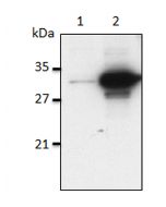
Method: IL-1α was analyzed by Western blot in cell extracts of bone marrow-derived dendritic cells (BMDCs) treated by LPS and the several inflammasome activators as indicated in the figure. Cell extracts were separated by SDS-PAGE under reducing conditions, transferred to nitrocellulose and incubated with anti-IL-1α (p18) (mouse), mAb (Teo-1) (1µg /ml). After addition of an anti-mouse secondary antibody coupled to HRP, proteins were visualized by a chemiluminescence detection system.
| Product Details | |
|---|---|
| Synonyms | Interleukin-1α IL-1alpha |
| Product Type | Monoclonal Antibody |
| Properties | |
| Clone | Teo-1 |
| Isotype | Mouse IgG |
| Source/Host | Purified from concentrated hybridoma tissue culture supernatant. |
| Immunogen/Antigen | Recombinant mouse mature IL-1α. |
| Application |
Western Blot: (1μg/ml) ELISA |
| Crossreactivity | Mouse |
| Specificity | Recognizes mouse IL-1α p18 cleaved and full-length fragments. |
| Purity | ≥95% (SDS-PAGE) |
| Purity Detail | Protein G-affinity purified. |
| Concentration | 1mg/ml |
| Formulation | Liquid. In PBS containing 10% glycerol and 0.02% sodium azide. |
| Shipping and Handling | |
| Shipping | BLUE ICE |
| Short Term Storage | +4°C |
| Long Term Storage | -20°C |
| Handling Advice |
After opening, prepare aliquots and store at -20°C. Avoid freeze/thaw cycles. |
| Use/Stability | Stable for at least 1 year after receipt when stored at -20°C. |
| Documents | |
| MSDS |
 Download PDF Download PDF |
| Product Specification Sheet | |
| Datasheet |
 Download PDF Download PDF |
The most prominent members of the interleukin-1 (IL-1) superfamily are IL-1α and IL-1β. They lack a signal peptide and are secreted by an unconventional, endoplasmic reticulum-Golgi-independent mechanism. IL-1α was reported to be more widely and constitutively expressed and has intracellular functions, but also acts locally in a membrane-bound form by activating IL-1R1. Additionally, passive release of IL-1α upon cell death can trigger a sterile inflammatory response to dying cells. The cleavage of IL-1α is not mediated by caspase-1 and is not required for binding to IL-1R1. Recently it has been observed that all activators of the inflammasome NLRP3/NALP3 induce the simultaneous secretion of IL-1α and IL-1β. Although most activators fully rely on the inflammasome for IL-1α secretion, some induce the processing and secretion of IL-1α in an inflammasome-independent manner.
- Formyl peptide receptor 1 signaling potentiates inflammatory brain injury: Z. Li, et al.; Sci. Transl. Med. 13, 605 (2021)


