Cookie Policy: This site uses cookies to improve your experience. You can find out more about our use of cookies in our Privacy Policy. By continuing to browse this site you agree to our use of cookies.
AdipoGen Life Sciences
anti-Periostin, mAb (Stiny-1)

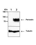
Whole tissue extracts from mouse liver (lane 1) or mammary tumor (lane 2) were separated by SDS-PAGE under reducing conditions, transferred to nitrocellulose and incubated with anti-Periostin, mAb (Stiny-1) (1μg/ml). Proteins were visualized by a chemiluminescence detection system.
Method: Normal Breast (A) or Breast Cancer (B) tissues were stained with Periostin, mAb (Stiny-1) (1:200) by standard immunohistochemistry.
| Product Details | |
|---|---|
| Synonyms | Osteoblast-specific Factor 2; OSF-2; Postn |
| Product Type | Monoclonal Antibody |
| Properties | |
| Clone | Stiny-1 |
| Isotype | Mouse IgG1κ |
| Source/Host | Purified from concentrated hybridoma tissue culture supernatant. |
| Immunogen/Antigen | Full-length human periostin. |
| Application |
ELISA |
| Crossreactivity |
Human Mouse |
| Specificity |
Recognizes human and mouse periostin. Detects the Fas1-4 domain (aa496-628) of human periostin. |
| Purity | ≥95% (SDS-PAGE) |
| Purity Detail | Protein G-affinity purified. |
| Concentration | 1mg/ml |
| Formulation | Liquid. In PBS containing 10% glycerol and 0.02% sodium azide. |
| Isotype Negative Control | |
| Shipping and Handling | |
| Shipping | BLUE ICE |
| Short Term Storage | +4°C |
| Long Term Storage | -20°C |
| Handling Advice |
After opening, prepare aliquots and store at -20°C. Avoid freeze/thaw cycles. |
| Use/Stability | Stable for at least 1 year after receipt when stored at -20°C. |
| Documents | |
| MSDS |
 Download PDF Download PDF |
| Product Specification Sheet | |
| Datasheet |
 Download PDF Download PDF |
Periostin is a 90-kDa matricellular protein that consists of a typical signal sequence, followed by a cysteine-rich region, an EMI domain (protein-protein interactions), four tandem fasciclin-like domains that are responsible for integrin binding, and a C-terminal region. Periostin was originally isolated as an osteoblast-specific factor that functions as a cell adhesion molecule for pre-osteoblasts and in osteoblast recruitment, attachment and spreading. Periostin is also involved in many fundamental biological processes such as cell proliferation, cell invasion and angiogenesis. Periostin expression is increased by both transforming growth factor β1 (TGF-β1) and bone morphogenetic protein 2 (BMP-2). Changes in periostin expression are commonly detected in various cancers and pre-cancerous conditions, and periostin may be involved in regulating cancer cell activities that contribute to tumorigenesis, cancer progression and metastasis. Periostin has shown to be involved in many aspects of allergic inflammation, such as eosinophil recruitment, airway remodeling, development of a Th2 phenotype and increased expression of inflammatory mediators. It is evaluated as a biomarker for bronchial asthma and airway inflammation.
- Interactions between cancer stem cells and their niche govern metastatic colonization: I. Malanchi, et al.; Nature 481, 85 (2012)
- Periostin contributes to the acquisition of multipotent stem cell-like properties in human mammary epithelial cells and breast cancer cells: X. Wang, et al.; PLoS One 8, e72962 (2013)
- The perivascular niche regulates breast tumour dormancy: C.M. Ghajar, et al.; Nat. Cell Biol. 15, 807-817 (2013)
- Matricellular protein periostin contributes to hepatic inflammation and fibrosis: Y. Huang, et al.; Am. J. Pathol. 185, 786 (2015)
- Endogenous semaphorin-7A impedes human lung fibroblast differentiation: S. Esnault, et al.; PLoS One 12, e0170207 (2017)
- IL-5-stimulated eosinophils adherent to periostin undergo stereotypic morphological changes and ADAM8-dependent migration: M.W. Johansson, et al.; Clin. Exp. Allergy 47, 1263 (2017)
- Periostin blockade overcomes chemoresistance via restricting the expansion of mesenchymal tumor subpopulations in breast cancer: Y. Nakazawa, et al., Sci. Rep. 8, (2018)
- Bone Marrow Mesenchymal Stromal Cell-Derived Periostin Promotes B-ALL Progression by Modulating CCL2 in Leukemia Cells: Z. Ma, et al.; Cell Rep. 26, 1533 (2019)
- Periostin Promotes Colorectal Tumorigenesis through Integrin-FAK-Src Pathway-Mediated YAP/TAZ Activation: H. Ma, et al.; Cell Rep. 30, 793 (2020) [IF, IHC]
- Periostin Short Fragment with Exon 17 via Aberrant Alternative Splicing Is Required for Breast Cancer Growth and Metastasis: Y. Ikeda-Iwabu, et al.; Cells 10, 892 (2021) [IF, IHC]
- Periostin deficiency reduces PD-1+ tumorassociated macrophage infiltration and enhances anti-PD-1 efficacy in colorectal cancer: T. Wei, et al.; Cell Rep. 42, 112090 (2023) [IF, IHC]
- Cancer-associated fibroblast-derived periostin promotes papillary thyroid tumor growth through integrin-FAK-STAT3 signaling: X. Jin, et al.; Theranostics 14, 3014 (2024) [IF, IHC]
- A Role for Periostin Pathological Variants and Their Interaction with HSP70-1a in Promoting Pancreatic Cancer Progression and Chemoresistance: Y. Tsunetoshi, et al.; Int. J. Mol. Sci. 25, 13205 (2024)


