Cookie Policy: This site uses cookies to improve your experience. You can find out more about our use of cookies in our Privacy Policy. By continuing to browse this site you agree to our use of cookies.
Chemodex
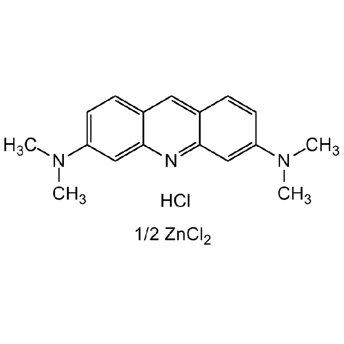
Acridine Orange hemi(zinc chloride)
As low as
81
CHF
CHF 81.00
In stock
Only %1 left
CDX-A0668-G02525 gCHF 81.00
CDX-A0668-G100100 gCHF 264.00


Specifications / Handling
| Product Details | |
|---|---|
| Synonyms | 3,6-Bis(dimethylamino)acridine hydrochloride zinc chloride double salt; Basic Orange 14; Euchrysin GNY; C.I. 46005; Acridine Orange |
| Product Type | Chemical |
| Properties | |
| Formula |
C17H20ClN3 . HCl . 1/2ZnCl2 |
| MW | 369.96 |
| CAS | 10127-02-3 |
| RTECS | AR7600000 |
| Source/Host Chemicals | Synthetic |
| Purity Chemicals | ≥90% (Dye content) |
| Appearance | Red to brown powder. |
| Solubility | Soluble in water (1 mg/ml) or ethanol (2mg/ml). |
| Identity | Determined by 1H-NMR. |
| Declaration | Manufactured by Chemodex. |
| Other Product Data |
Click here for Original Manufacturer Product Datasheet |
| InChi Key | VADJQOXWNSPOQA-UHFFFAOYSA-L |
| Smiles | Cl[Zn]Cl.CN(C)C1=CC=C2C(N=C(C=C(N(C)C)C=C3)C3=C2)=C1.Cl |
| Shipping and Handling | |
| Shipping | AMBIENT |
| Short Term Storage | +20°C |
| Long Term Storage | +20°C |
| Handling Advice |
Keep under inert gas. Very hygroscopic. |
| Use/Stability | Stable for at least 2 years after receipt when stored at RT. |
| Documents | |
| Product Specification Sheet | |
| Datasheet |
 Download PDF Download PDF |
Scientific Background Information
Product Description
Acridine Orange hemi(zinc chloride) salt is a cell-permeable metachromatic fluorescent dye that stains DNA and RNA. It is used as a nucleic acid-selective fluorescent cationic dye useful for cell cycle determination. Being cell-permeable, it interacts with DNA and RNA by intercalation or electrostatic attractions respectively. When bound to DNA, it is very similar spectrally to fluorescein, with an excitation maximum at 502nm and an emission maximum at 525nm (green). When acridine orange associates with RNA, the excitation maximum shifts to 460nm (blue), and the emission maximum shifts to 650nm (red). Acridine orange will also enter acidic compartments such as lysosomes where it becomes protonated and sequestered. Within these low pH vesicles, the dye emits red fluorescence when excited by blue light. Thus, acridine orange can be used to visualize primary lysosomes and phagolysosomes that may include products of phagocytosis of apoptotic cells. The dye is often used in epifluorescence microscopy and flow cytometry.It allows for visual detection of nucleic acids on agarose and polyacrylamide gels, can be used for differentiation of dsDNA (green fluorescence) and ssDNA/RNA (orange fluorescence) and as a vitality test for determination of living cells. Spectral data: λEx=502nm, λEm=525nm (green, double strands) / λEx=460nm, λEm=650nm (red, single strands).
Product-specific References
(1) F.H. Kasten; Int. Rev. Cytol. 21, 141 (1967) (Review) | (2) J.F. Golden & S.S. West; J. Histochem. Cytochem. 22, 495 (1974) | (3) J.F. Golden, et al.; J. Histochem. Cytochem. 27, 522 (1979) | (4) J.K. Frost, et al.; J. Histochem. Cytochem. 27, 545 (1979) | (5) H.W. Tyrer, et al.; J. Histochem. Cytochem. 27, 552 (1979) | (6) R.N. Paul; Stain Technol. 55, 195 (1980) | (7) S. Mirrett; Inf. Contr. Hosp. Epidemiol. 3, 250 (1982) (Review) | (8) Z. Darzynkiewicz, et al.; Curr. Protoc. Cytom. Chapter 7, Unit 7.3 (2004) (Review) | (9) R.W. Sabnis; Handbook of Fluorescent Dyes and Probes (2015)