Cookie Policy: This site uses cookies to improve your experience. You can find out more about our use of cookies in our Privacy Policy. By continuing to browse this site you agree to our use of cookies.
Chemodex
Hoechst 33342 Solution
As low as
135
CHF
CHF 135.00
In stock
Only %1 left
CDX-B0447-L0055 mlCHF 135.00


Specifications / Handling
| Product Details | |
|---|---|
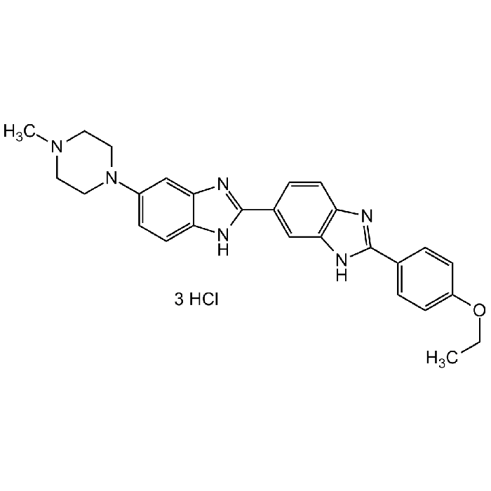
| Synonyms | 2'-(4-Ethoxyphenyl)-5-(4-methyl-1-piperazinyl)-2,5'-bi-1H-benzimidazole trihydrochloride; BisBenzimide H 33342; HOE 33342 |
| Product Type | Chemical |
| Properties | |
| Formula |
C27H28N6O . 3HCl . xH2O |
| MW | 561.93 |
| CAS | 23491-52-3 |
| RTECS | DT4200000 |
| Source/Host Chemicals | Synthetic |
| Purity Chemicals | ≥98% (HPLC) |
| Appearance | Liquid. |
| Solubility | Soluble in water. |
| Concentration | 1mg/ml in water |
| Identity | Determined by 1H-NMR. |
| Declaration | Manufactured by Chemodex. |
| Other Product Data |
Click here for Original Manufacturer Product Datasheet |
| InChi Key | PRDFBSVERLRRMY-UHFFFAOYSA-N |
| Smiles | CN(CC1)CCN1C2=CC=C(NC(C3=CC(NC(C4=CC=C(OCC)C=C4)=N5)=C5C=C3)=N6)C6=C2 |
| Shipping and Handling | |
| Shipping | AMBIENT |
| Short Term Storage | +4°C |
| Long Term Storage | +4°C |
| Handling Advice | Protect from light and moisture. |
| Use/Stability | Stable for at least 2 years after receipt when stored at +4°C. |
| Documents | |
| MSDS |
 Download PDF Download PDF |
| Product Specification Sheet | |
| Datasheet |
Download PDF |
Scientific Background Information
Product Description
The Hoechst stains are a family of fluorescent stains for labeling DNA in fluorescence microscopy. The blue fluorescent Hoechst dye is a cell permeable nucleic acid stain that has multiple applications, including sensitive detection of DNA in the presence of RNA in agarose gels, automated DNA determination, sensitive determination of cell number and chromosome sorting. Useful vital stain for the flow cytometric recognition of DNA damage and other viability measurements by monitoring the emission spectral shifts of the dyes. Because this fluorescent stain labels DNA, it can also be used to visualize nuclei and mitochondria. Hoechst 33342 is a cell-permeable, benzimidazole dye that stains DNA by binding to the minor groove of adenine and thymine-rich sequences. It emits blue fluorescence (excitation 350 nm/emission maximum 461 nm) when bound to double stranded DNA and is useful as a marker of nuclei for cell cycle studies and to distinguish nuclear morphology in apoptotic cells.
Product-specific References
(1) D.J. Pierce et al.: Exp. Hematol. 35(9), 1437 (2007) | (2) M.E. Lalande et al.: Proc. Natl. Acad. Sci. USA 78, 363 (1981) | (3) M.J. Lydon et al.: J. Cell. Physiol. 102, 175 (1980)