Cookie Policy: This site uses cookies to improve your experience. You can find out more about our use of cookies in our Privacy Policy. By continuing to browse this site you agree to our use of cookies.
Chemodex
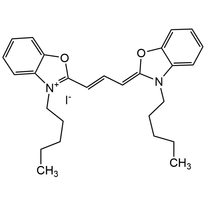
3,3'-Dipentyloxacarbocyanine iodide
As low as
116
CHF
CHF 116.00
In stock
Only %1 left
CDX-D0128-M05050 mgCHF 116.00
CDX-D0128-M100100 mgCHF 200.00
CDX-D0128-M250250 mgCHF 400.00


Specifications / Handling
| Product Details | |
|---|---|
| Synonyms | DiOC5(3); NK 2453 |
| Product Type | Chemical |
| Properties | |
| Formula | C27H33IN2O2 |
| MW | 544.47 |
| CAS | 53213-81-3 |
| Source/Host Chemicals | Synthetic |
| Purity Chemicals | ≥98% (HPLC) |
| Appearance | Coral powder. |
| Solubility | Soluble in DMSO or DMF. |
| Identity | Determined by 1H-NMR. |
| Declaration | Manufactured by Chemodex. |
| Other Product Data |
Click here for Original Manufacturer Product Datasheet |
| InChi Key | CDLMLYASVIQVPH-UHFFFAOYSA-M |
| Smiles | C(CCCC)N1C=2C(OC1=CC=CC3=[N+](CCCCC)C=4C(O3)=CC=CC4)=CC=CC2.[I-] |
| Shipping and Handling | |
| Shipping | AMBIENT |
| Short Term Storage | +4°C |
| Long Term Storage | -20°C |
| Handling Advice |
Keep under inert gas. Protect from light and oxygen. |
| Use/Stability | Stable for at least 2 years after receipt when stored at -20°C. |
| Documents | |
| Product Specification Sheet | |
| Datasheet |
 Download PDF Download PDF |
Scientific Background Information
Product Description
3,3'-Dipentyloxacarbocyanine iodide is a lipophilic carbocyanine dye used primarily for membrane potential studies and cell labeling. Used to monitor mitochondrial or plasma membrane potential in live cells. The dye accumulates in polarized membranes and fluorescence intensity correlates with membrane potential. The dye can label lipid membranes due to its hydrophobic side chains and us useful for flow cytometry or fluorescence microscopy. Spectral data: λex 484 nm, λem 509 nm in methanol. Intermediate for the synthesis of fluorescent probes.
Product-specific References
(1) G. Meissner; J. Biol. Chem. 256, 636 (1981) | (2) J.G. Monroe & J.C. Cambier; J. Immunol. 131, 2641 (1983) | (3) R.L. Gallo, et al.; Arch. Biochem. Biophys. 235, 544 (1984) | (4) H.A. Crissman, et al.; Exp. Cell Res. 174, 388 (1988) | (5) H.A. Crissman, et al.; Methods Cell Biol. 33, 89 (1990) | (6) M. Yamashita, et al.; FEBS J. 273, 3585 (2006) | (7) B. Zhang, et al.; Biomaterials 52, 14 (2015)