Cookie Policy: This site uses cookies to improve your experience. You can find out more about our use of cookies in our Privacy Policy. By continuing to browse this site you agree to our use of cookies.
Chemodex
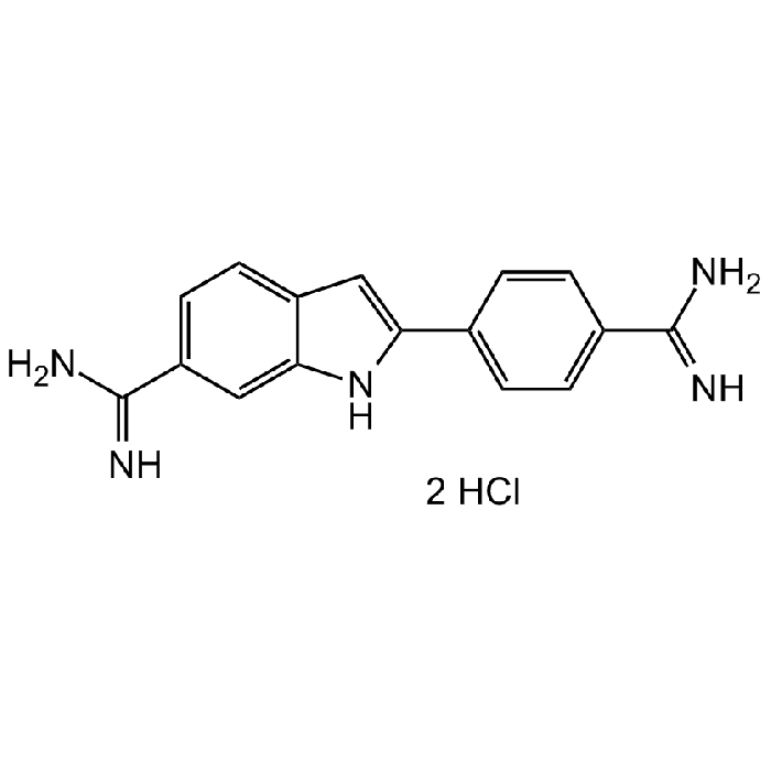
DAPI dihydrochloride Solution (10 mM in water)

| Product Details | |
|---|---|
| Synonyms | 2-(4-Amidinophenyl)-6-indolecarbamidine dihydrochloride; 4',6-Diamidino-2-phenylindole dihydrochloride; FxCycle Violet |
| Product Type | Chemical |
| Properties | |
| Formula | C16H15N5 . 2HCl |
| MW | 350.25 |
| CAS | 28718-90-3 |
| Purity Chemicals | ≥95% (HPLC) |
| Appearance | Liquid. |
| Solubility | Soluble in water. |
| Concentration | 10mM in water |
| Identity | Determined by 1H-NMR. |
| Declaration | Manufactured by Chemodex. |
| Other Product Data |
Click here for Original Manufacturer Product Datasheet |
| InChi Key | FPNZBYLXNYPRLR-UHFFFAOYSA-N |
| Smiles | NC(C1=CC=C2C(NC(C3=CC=C(C(N)=N)C=C3)=C2)=C1)=N.Cl.Cl |
| Shipping and Handling | |
| Shipping | AMBIENT |
| Short Term Storage | -20°C |
| Long Term Storage | -20°C |
| Handling Advice | Protect from light and moisture. |
| Use/Stability | Stable for at least 2 years after receipt when stored at -20°C. |
| Documents | |
| Product Specification Sheet | |
| Datasheet |
 Download PDF Download PDF |
DAPI (4,6-Diamidino-2-phenylindole dihydrochloride) is a cell permeable, fluorescent dye that is a minor groove-binding probe for DNA. Binds to the minor groove of double-stranded DNA (preferentially to AT rich DNA), forming a stable complex which fluoresces approximately 20 times greater than DAPI alone. DAPI is several times more sensitive than ethidium bromide for staining DNA in agarose gels. It may be used for photofootprinting of DNA, to detect annealed probes in blotting applications by specifically visualizing the double-stranded complex, and to study the changes in DNA and analyze DNA content during apoptosis using flow cytometry. DAPI staining has also been shown to be a sensitive and specific detection method for mycoplasma. Spectral Data: Excitation: λex 340 nm; Emission: λem 488 nm (only DAPI). Excitation: λex 360nm; Emission: λem 460nm (DAPI-DNA complex).
(1) W.C. Russell, et al.; Nature 253, 461 (1975) | (2) I.W. Taylor & B.K. Milthorpe; J. Histochem. Cytochem. 28, 1224 (1980) | (3) F. Otto & K.C. Tsou; Stain Technol. 60, 7 (1985) | (4) C. Cubria, et al.; Comp. Biochem. Physiol. C. 105, 251 (1994) | (5) M.A. Hotz, et al.; Cytometry 15, 237 (1994) | (6) J. Kapuscinski; Biotech. Histochem. 70, 220 (1995) | (7) E. Buel & M. Schwartz; J. Forensic Sci. 40, 275 (1995) | (8) D. Zink, et al.; Methods: A Companion to Methods in Enzymology 29, 42 (2003) | (9) L. Rodgers; CSH Protoc. 2006, pdb.prot4438 (2006) | (10) B. Chazotte; CSH Protoc. 2011, pdb.prot5556 (2011) | (11) A. Biancardi, et al.; Phys. Chem. Chem. Phys. 15, 4596 (2013) | (12) M. Jez, et al.; Histochem. Cell Biol. 139, 195 (2013) | (13) G. Yahav, et al.; Cytometry A 89, 644 (2016)


