Cookie Policy: This site uses cookies to improve your experience. You can find out more about our use of cookies in our Privacy Policy. By continuing to browse this site you agree to our use of cookies.
Chemodex
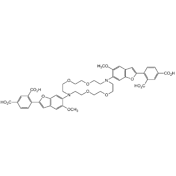
PBFI

| Product Details | |
|---|---|
| Synonyms | Potassium-binding Benzofuran Isophthalate |
| Product Type | Chemical |
| Properties | |
| Formula |
C46H46N2O16 |
| MW | 882.86 |
| CAS | 124549-11-7 |
| Purity Chemicals | ~80% (HPLC) |
| Appearance | Brownish-yellow powder. |
| Solubility | Soluble in methanol or DMSO. |
| Declaration | Manufactured by Chemodex. |
| Other Product Data |
Click here for Original Manufacturer Product Datasheet |
| InChi Key | YOQMJMHTHWYNIO-UHFFFAOYSA-N |
| Smiles | COC1=CC2=C(OC(C3=C(C(O)=O)C=C(C(O)=O)C=C3)=C2)C=C1N4CCOCCOCCN(C5=CC(OC(C6=C(C(O)=O)C=C(C(O)=O)C=C6)=C7)=C7C=C5OC)CCOCCOCC4 |
| Shipping and Handling | |
| Shipping | AMBIENT |
| Short Term Storage | -20°C |
| Long Term Storage | -20°C |
| Handling Advice | Protect from light and moisture. |
| Use/Stability | Stable for at least 2 years after receipt when stored at -20°C. |
| Documents | |
| Product Specification Sheet | |
| Datasheet |
 Download PDF Download PDF |
PBFI is a cell-impermeant fluorescent indicator for potassium ions, suitable for detection of physiological concentrations of K+ in the presence of other monovalent cations. PBFI is used to measure intracellular potassium (K+) fluxes in animal cells and in plant cells and vacuoles. The observation that intracellular K+ levels are a controlling factor in apoptotic cell death pathways, increased the interest for PBFI. PBFI has been used for detecting adrenoceptor-stimulated decreases of intracellular K+ concentration in astrocytes and neurons, evaluating the mediating effects of K+ depletion on monocytic cell necrosis, measuring intracellular K+ fluxes associated with apoptotic cell shrinkage, monitoring mitochondrial KATP channel activation, or detecting elevated intracellular K+ levels associated with HIV-induced cytopathology. Flow cytometric measurements using UV argon-ion laser excitation (351nm and 364nm) of PBFI indicate that K+ efflux induces shrinkage of apoptotic cells and is a trigger for caspase activation. Spectral data: λex 336nm;, λem 557nm.
(1) P. Jezek, et al.; J. Biol. Chem. 265, 10522 (1990) | (2) S.E. Kasner & M.B. Ganz; Am. J. Physiol. 262, F462 (1992) | (3) K. Venema, et al., Biochim. Biophys. Acta 1146, 87 (1993) | (4) R. Crossley, et al.; J. Chem. Soc. Perk. Trans. 2, 513 (1994) | (5) K. Meuwis; Biophys. J. 68, 2469 (1995) | (6) H. Muyderman, et al.; Neurochem. Int. 38, 269 (2001) | (7) A.L. Cook, et al.; Cell Signal. 14, 1023 (2002) | (8) S.J. Halperin & J.P. Lynch; J. Exp. Bot. 54, 2035 (2003) | (9) D. Liu, et al.; J. Neurochem. 86, 966 (2003) | (10) A.D. Costa, et al.; Am. J. Physiol. Heart Circ. Physiol. 290, H406 (2006) | (11) P. Andersson, et al.; Toxicol. In Vitro 20, 986 (2006) | (12) S. Jorgensen, et al.; Methods Mol. Biol. 491, 257 (2008) | (13) R.W. Sabnis; Handbook of biological dyes and stains (2010) | (14) C.S. Arlehamn, et al.; J. Biol. Chem. 285, 10508 (2010)


