Cookie Policy: This site uses cookies to improve your experience. You can find out more about our use of cookies in our Privacy Policy. By continuing to browse this site you agree to our use of cookies.
AdipoGen Life Sciences
anti-PD-1 (human), mAb (AG-IHC001)
As low as
220
CHF
CHF 220.00
In stock
Only %1 left
AG-20B-6020-R100100 µlCHF 220.00


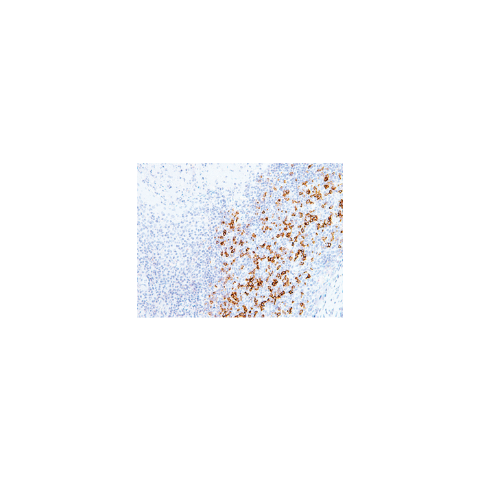
Figure 1: Immunohistochemical staining of PD-1 using anti-PD-1 (human), mAb (AG-IHC001) (Prod. No. AG-20B-6020) in formalin-fixed and paraffin-embedded (FFPE) human tonsil tissue.
Specifications / Handling
| Product Details | |
|---|---|
| Synonyms | CD279; Programmed Cell Death Protein 1 |
| Product Type | Monoclonal Antibody |
| Properties | |
| Clone | AG-IHC001 |
| Isotype | Mouse IgG2b |
| Source/Host | Purified from concentrated hybridoma tissue culture supernatant. |
| Immunogen/Antigen | Recombinant human PD-1 fused to a His-tag. |
| Application |
Immunohistochemistry: 1:100 - 1:200 for formalin-fixed, paraffin-embedded tissue sections (frozen sections not validated). Recommended Positive Control: Tonsil or Lymph Node |
| Crossreactivity | Human |
| Specificity |
Recognizes human PD-1 (CD279). |
| Purity | Protein A/G purified. |
| Formulation | Liquid. In Tris Buffer, pH 7.4, containing 1% BSA and <0.1% sodium azide. |
| Isotype Negative Control | |
| Other Product Data |
UniProt Link Q15116: PD-1 (human) |
| Accession Number | Q15116 |
| Shipping and Handling | |
| Shipping | BLUE ICE |
| Short Term Storage | +4°C |
| Long Term Storage | +4°C |
| Handling Advice | Do not freeze. |
| Use/Stability | Stable for at least 1 year after receipt when stored at +4°C. |
| Documents | |
| Protocols |
 Download PDF Download PDF |
| MSDS |
Download PDF |
| Product Specification Sheet | |
| Datasheet |
Download PDF |
Scientific Background Information
Product Description
Programmed Death 1 (PD-1) is a member of the CD28/CTLA-4 family of T cell regulators, expressed as a co-receptor on the surface of activated T cells, B cells and macrophages. Several studies have suggested that the PD-1/PD-L1 signaling pathway may be linked to antitumor immunity, as PD-L1 has been shown to induce apoptosis of activated T cells or inhibit activity of cytotoxic T cells. In comparison to CD10 and Bcl-6, PD-1 is expressed by fewer B cells and has therefore been considered a more specific and useful diagnostic marker for angioimmunoblastic T cell lymphoma. Therapies targeted toward the PD-1 receptor have shown remarkable clinical responses in patients with various types of cancer, including non-small-cell lung cancer, melanoma and renal-cell cancer.
This antibody is intended to qualitatively identify by light microscopy the presence of associated antigens in sections of formalin-fixed, paraffin-embedded tissue sections using IHC test methods. It has been optimized and validated using the BOND-MAX fully automated IHC&ISH stainer (see Protocol).
Related Products
-
![CD279 [PD-1] (mouse):Fc (human) (rec.)](https://adipogen.com/media/catalog/product/placeholder/default/adipogen_logo_bw_3.png) CD279 [PD-1] (mouse):Fc (human) (rec.) CHI-MF-111PD1
CD279 [PD-1] (mouse):Fc (human) (rec.) CHI-MF-111PD1 -
CD279 [PD-1] (mouse):Fc (mouse) (rec.) CHI-MF-110PD1
-
CD279 [PD-1] (human):Fc (mouse) (rec.) CHI-HF-211PD1
-
CD279 [PD-1] (human):Fc (human) (rec.) CHI-HF-210PD1
-
CD279 [PD-1] (human) (rec.) (His) CHI-HF-201PD1
-
CD279 [PD-1] (human) (rec.) CHI-HF-200PD1
-
CD279 [PD-1] (human)-muIg Fusion Protein ANC-549-020.
-
anti-CD279 [PD-1] (human), mAb (ANC4H6) ANC-279-020.