Cookie Policy: This site uses cookies to improve your experience. You can find out more about our use of cookies in our Privacy Policy. By continuing to browse this site you agree to our use of cookies.
Antibodies
Toggle Nav
Items 521-530 of 874
-
-
-
-
-
-

-
 anti-IgG1 (mouse), Rabbit Monoclonal (RM106) (Biotin)
REV-31-1002-02 REV-31-1002-02-C050 50 µg CHF 468.00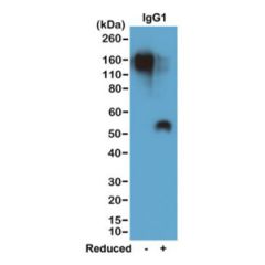
-

-
anti-IgG3 (mouse), Rabbit Monoclonal (RM218) (Biotin)
REV-31-1094-02 REV-31-1094-02-C050 50 µg CHF 468.00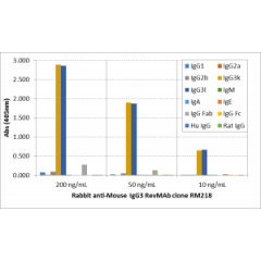
-
Items 521-530 of 874
Shop By
Active Filters
Search Filter
- Application WB Remove This Item
Product Type
Product Line / Research Area
- Antibody 873 items
- Immunohistochemistry 242 items
- Apoptosis 47 items
- Angiogenesis 40 items
- Autophagy 2 items
- Bone Research 3 items
- Cancer 506 items
- Cell Cycle 17 items
- Cell Death 32 items
- Cytokines 14 items
- Cytoskeleton 43 items
- Epigenetics 80 items
- Immunology 645 items
- Immunometabolism 16 items
- Immuno-Oncology 15 items
- Inflammasomes 28 items
- Inflammation 380 items
- Innate Immunity 53 items
- Metabolism 152 items
- Mitochondria 4 items
- Neurobiology 33 items
- Neurodegenerative Disease 9 items
- Nitric Oxide 1 item
- Obesity 18 items
- Oxidative Stress 3 items
- Pathology 238 items
- Posttranslational Modifications 87 items
- Stem Cell Research 20 items
- TNF Superfamily 23 items
- Ubiquitin Pathway 4 items
Crossreactivity
- All 29 items
- Avian 1 item
- Bovine 3 items
- Chicken 1 item
- Ciliates 1 item
- Cow 1 item
- Dog 13 items
- Drosophila 3 items
- Fungus 1 item
- Goat 2 items
- Guinea pig 1 item
- Hamster 1 item
- HCV 2 items
- Horse 1 item
- Human 658 items
- Listeria monocytogenes 2 items
- Listeria sp. 1 item
- Listeria spp. 1 item
- Monkey 12 items
- Mouse 156 items
- Pig 12 items
- Primate 24 items
- Rabbit 4 items
- Rat 60 items
- Sheep 1 item
- Virus 8 items
- Zebra fish 2 items
- All Vertebrates 66 items
- Pseudomonas aeruginosa 1 item
- Legionella pneumophila 1 item
- Clostridioides difficile 3 items
Brand
Isotype
- Goat IgG 2 items
- Hamster IgG 7 items
- Human IgG1 16 items
- Human IgG1κ 4 items
- Human IgG2λ 7 items
- IgG 1 item
- IgG1 5 items
- IgG2a 2 items
- IgG2b 1 item
- Mouse IgG 2 items
- Mouse IgG1 116 items
- Mouse IgG1κ 96 items
- Mouse IgG2a 37 items
- Mouse IgG2aκ 16 items
- Mouse IgG2b 7 items
- Mouse IgG2bκ 13 items
- Mouse IgG2bκ. 1 item
- Mouse IgG2bλ 1 item
- Mouse IgG3 1 item
- Mouse IgG3κ 10 items
- Mouse IgM 4 items
- Mouse IgMκ 3 items
- Mouse IgG2bκ 1 item
- Rabbit IgG 322 items
- Rabbit/Human Chimeric<br />Rabbit Fv/ Human IgG CH and Cκ 2 items
- Rat IgG1 1 item
- Rat IgG1κ 8 items
- Rat IgG2a 1 item
- Rat IgG2aκ 17 items
Clone
- 1.3.3.22 5 items
- 1.G11B1 5 items
- 10.1 8 items
- 10B12 1 item
- 10H 1 item
- 112A1021 1 item
- 117C 1 item
- 11B4 1 item
- 11D3 1 item
- 11E3 2 items
- 11F9 1 item
- 12G6 1 item
- 13F11 1 item
- 145-2C11 6 items
- 15.2 5 items
- 169-1-E4.3 4 items
- 17A9 1 item
- 17B4 2 items
- 18H2 1 item
- 18H5 1 item
- 1B6 1 item
- 1C3 1 item
- 1C4 2 items
- 1E7E8 5 items
- 1G12 1 item
- 24-31 5 items
- 256-E 1 item
- 2A12 1 item
- 2B8 1 item
- 2E12 1 item
- 3(4H3) 1 item
- 3(6D3) 1 item
- 38.0 5 items
- 3A5 1 item
- 3B10 2 items
- 3C 1 item
- 3C5 1 item
- 3F7B6 5 items
- 41B436 1 item
- 43A1 6 items
- 4B7R 5 items
- 4E11 1 item
- 5B-3B1 1 item
- 5F12 1 item
- 5G10 1 item
- 5H11 1 item
- 6A8 1 item
- 6D8 1 item
- 7B10 1 item
- 7H11 1 item
- 8A5 1 item
- 9G10 1 item
- AB66-6-C10 1 item
- AB68-A09 2 items
- AB75-C03 1 item
- AB79-E11 1 item
- AB83-C12 1 item
- AC18F 2 items
- AC384 3 items
- Adri-1 1 item
- AG103 1 item
- Alme-1 2 items
- ANC5D6 4 items
- APO-1-3 1 item
- AXO45 1 item
- Bally-1 2 items
- Bamboo-1 1 item
- Bcl-2/100 4 items
- Birdy-1 1 item
- Birdy-2 1 item
- BPC 4 3 items
- BQ16 5 items
- BRA-10G 5 items
- BU33 5 items
- BU38 5 items
- BU52 1 item
- BU61 5 items
- BU75 5 items
- C15 1 item
- Carly-1-4 1 item
- Casper-1 2 items
- Casper-2 2 items
- Clint-1 1 item
- Covi-1 1 item
- Covi-2 1 item
- CR213-2AG 1 item
- Cryo-1 1 item
- Cryo-2 2 items
- D1L165-6 1 item
- D1L357-1-4 1 item
- Dave-2 1 item
- DFT1 5 items
- DL86-3AG 1 item
- E11 5 items
- EIC 1 item
- Eowyn-1 1 item
- ERIC-1 3 items
- EWC 1 item
- EWF 1 item
- F2C 1 item
- FG224-7 1 item
- FG322-3 1 item
- FG369-1 1 item
- FG98-6 1 item
- Flamy-1 2 items
- FT62-6 1 item
- FT86-4 1 item
- Giby-1-4 2 items
- Giusepi-1-4 1 item
- GT335 2 items
- HADI 773 1 item
- Hely-1 1 item
- HNEJ-2 2 items
- HRB 149 1 item
- HS501 1 item
- hu5c8 1 item
- Huf 5.4 2 items
- I 178G 1 item
- ID 177 1 item
- IE273 1 item
- IL33026B 1 item
- IL33068A 1 item
- IL33305B 1 item
- IST-9 1 item
- J1D9 4 items
- J1G53-3 1 item
- Jacky-1 1 item
- JCM-1 1 item
- Jussy-1 1 item
- Kairos 108-4 1 item
- Kairos 397-7 1 item
- Kairos-1 1 item
- Kairos-37 1 item
- Kairos4-153AD 1 item
- Kairos4-397G 1 item
- Lecty-1 1 item
- Lise-1 1 item
- M4173 1 item
- MADI 04 1 item
- MADI 1147 1 item
- MF333F 1 item
- MOPC31C 6 items
- MRB 46L 1 item
- MREL 127 1 item
- MRES 06 1 item
- MS112-IIA1 1 item
- Nady-1 1 item
- Naipa-1 1 item
- Nely-1 1 item
- NF6 2 items
- Nhat-1 1 item
- OMNI379 2 items
- p6007 1 item
- p6017 1 item
- Pab240 3 items
- PF105B 1 item
- PF13-3 1 item
- PF183E 1 item
- PF299-1 1 item
- PHK121-H2 1 item
- RADI 264 1 item
- RM101 1 item
- RM102 1 item
- RM105 2 items
- RM106 2 items
- RM111 1 item
- RM112 2 items
- RM113 2 items
- RM114 2 items
- RM120 2 items
- RM130 1 item
- RM135 1 item
- RM137 1 item
- RM140 1 item
- RM141 1 item
- RM146 2 items
- RM146X 1 item
- RM147 1 item
- RM149 1 item
- RM150 1 item
- RM151 1 item
- RM154 1 item
- RM155 1 item
- RM156 1 item
- RM157 1 item
- RM159 1 item
- RM160 1 item
- RM161 1 item
- RM162 1 item
- RM163 1 item
- RM164 1 item
- RM165 1 item
- RM166 1 item
- RM167 1 item
- RM168 1 item
- RM169 1 item
- RM171 1 item
- RM172 1 item
- RM175 1 item
- RM179 1 item
- RM180 1 item
- RM181 1 item
- RM186 1 item
- RM188 1 item
- RM190 1 item
- RM191 2 items
- RM192 2 items
- RM193 2 items
- RM194 1 item
- RM195 1 item
- RM199 1 item
- RM201 1 item
- RM202 1 item
- RM204 1 item
- RM205 1 item
- RM208 1 item
- RM212 1 item
- RM214 1 item
- RM215 1 item
- RM216 1 item
- RM218 2 items
- RM221 1 item
- RM222 1 item
- RM224 1 item
- RM225 1 item
- RM226 1 item
- RM228 1 item
- RM230 1 item
- RM232 1 item
- RM233 1 item
- RM234 1 item
- RM235 1 item
- RM238 1 item
- RM240 2 items
- RM241 1 item
- RM242 1 item
- RM243 1 item
- RM244 1 item
- RM245 1 item
- RM246 1 item
- RM247 1 item
- RM248 1 item
- RM249 1 item
- RM250 1 item
- RM251 1 item
- RM252 1 item
- RM253 1 item
- RM254 1 item
- RM255 1 item
- RM256 1 item
- RM257 1 item
- RM258 1 item
- RM259 1 item
- RM260 1 item
- RM261 1 item
- RM262 1 item
- RM263 2 items
- RM264 1 item
- RM265 1 item
- RM266 1 item
- RM267 1 item
- RM268 1 item
- RM269 1 item
- RM270 1 item
- RM271 1 item
- RM272 1 item
- RM273 1 item
- RM274 1 item
- RM275 1 item
- RM276 1 item
- RM277 1 item
- RM278 1 item
- RM279 1 item
- RM280 1 item
- RM281 1 item
- RM283 1 item
- RM284 1 item
- RM285 1 item
- RM287 1 item
- RM288 1 item
- RM289 1 item
- RM290 1 item
- RM291 1 item
- RM292 1 item
- RM293 1 item
- RM294 1 item
- RM296 1 item
- RM297 1 item
- RM298 1 item
- RM299 1 item
- RM300 1 item
- RM301 1 item
- RM302 1 item
- RM303 1 item
- RM304 1 item
- RM305 2 items
- RM306 1 item
- RM307 2 items
- RM308 1 item
- RM309 1 item
- RM310 1 item
- RM312 1 item
- RM313 1 item
- RM314 1 item
- RM315 1 item
- RM316 1 item
- RM317 1 item
- RM318 1 item
- RM320 1 item
- RM322 1 item
- RM323 2 items
- RM324 1 item
- RM325 1 item
- RM326 1 item
- RM327 1 item
- RM328 1 item
- RM330 1 item
- RM331 1 item
- RM332 1 item
- RM333 1 item
- RM334 1 item
- RM335 1 item
- RM336 2 items
- RM337 1 item
- RM339 1 item
- RM341 1 item
- RM344 1 item
- RM345 1 item
- RM346 1 item
- RM347 1 item
- RM348 1 item
- RM349 1 item
- RM350 1 item
- RM351 1 item
- RM352 1 item
- RM353 1 item
- RM354 1 item
- RM355 1 item
- RM356 1 item
- RM357 1 item
- RM359 1 item
- RM360 1 item
- RM361 1 item
- RM363 2 items
- RM364 1 item
- RM365 1 item
- RM366 1 item
- RM367 1 item
- RM368 1 item
- RM369 1 item
- RM370 1 item
- RM371 1 item
- RM372 1 item
- RM373 1 item
- RM374 1 item
- RM375 1 item
- RM376 1 item
- RM377 1 item
- RM378 1 item
- RM379 1 item
- RM381 1 item
- RM382 1 item
- RM383 1 item
- RM384 1 item
- RM385 1 item
- RM386 1 item
- RM387 1 item
- RM388 1 item
- RM389 1 item
- RM391 1 item
- RM393 1 item
- RM394 1 item
- RM395 1 item
- RM396 1 item
- RM397 1 item
- RM398 1 item
- RM399 1 item
- RM401 1 item
- RM402 1 item
- RM403 1 item
- RM404 1 item
- RM405 1 item
- RM406 1 item
- RM407 2 items
- RM408 1 item
- RM409 1 item
- RM415 1 item
- RM416 1 item
- RM419 2 items
- RM422 1 item
- RM423 1 item
- RM424 1 item
- RM425 1 item
- RM427 1 item
- RM431 1 item
- RM432 1 item
- RM433 1 item
- RM437 1 item
- RM438 1 item
- RM439 1 item
- RM442 1 item
- RM443 1 item
- RM444 1 item
- RM445 1 item
- RM446 1 item
- RM447 1 item
- RM448 1 item
- RM449 1 item
- RM451 1 item
- RM452 1 item
- RM453 1 item
- RM454 1 item
- RM455 1 item
- RM456 1 item
- RM457 1 item
- RM458 1 item
- RM459 1 item
- RM461 1 item
- RM463 1 item
- RM464 1 item
- RM465 1 item
- RM468 1 item
- RM469 1 item
- RM470 1 item
- RM471 1 item
- RM472 1 item
- RM473 1 item
- RM478 1 item
- RM482 1 item
- RM483 1 item
- RM484 1 item
- RM485 1 item
- RM486 1 item
- RM487 1 item
- RM488 1 item
- RM489 1 item
- RM490 1 item
- RM492 1 item
- RM493 1 item
- RM494 1 item
- RM496 1 item
- RM497 1 item
- RM498 1 item
- RM499 1 item
- RM500 1 item
- RM501 1 item
- RM502 1 item
- RM503 1 item
- RM504 1 item
- RM505 1 item
- RM506 1 item
- RM507 1 item
- RM514 1 item
- RM515 1 item
- RM516 1 item
- RM518 1 item
- RM519 1 item
- RM520 1 item
- RM521 1 item
- RM7 2 items
- RM8 2 items
- RMG03 2 items
- RMH01 1 item
- RMH02 1 item
- RP159-1 1 item
- RREL 803 1 item
- RRES 07 1 item
- RTD4E10 1 item
- S11B 1 item
- S2R233-1 1 item
- S6R82-2 1 item
- Serpy-1-4 1 item
- SF9 1 item
- SH1177-F3 1 item
- SH1352-B11 1 item
- SH1352-G9 1 item
- SH1429-B1 1 item
- SH1579-B7 1 item
- SH313-B5 1 item
- SIPC 3385 3 items
- Skiny-1 1 item
- SN5c 5 items
- SN8 5 items
- ST33868 1 item
- Stiny-1 3 items
- TDR31.1 6 items
- tek16 1 item
- tek2 1 item
- tek9 1 item
- Teo-1 1 item
- TEPC 183 3 items
- TEU318 1 item
- TI 339H 1 item
- TOB5-D4 1 item
- TUN219-2C1 (Hypermyc) – 9E10 improved variant 3 items
- UCHT1 8 items
- UCHT4 5 items
- UM-8D6 6 items
- UM4D4 4 items
- UMA9 5 items
- VIF090-A6 1 item
- VIF137-E7 1 item
- VIF140-A1 1 item
- VP63 1 item
- Zagy-1 1 item
- Zagy-2 1 item
- Zippy-1 1 item