Cookie Policy: This site uses cookies to improve your experience. You can find out more about our use of cookies in our Privacy Policy. By continuing to browse this site you agree to our use of cookies.
Immunology
-
 anti-5-Carboxylcytosine (5-caC), Rabbit Monoclonal (RM462)
REV-31-1354-00 REV-31-1354-00-C050 50 µg CHF 623.00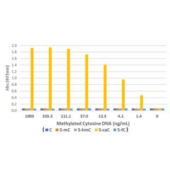
-
anti-5-Formylcytosines (5-fC), Rabbit Monoclonal (RM477)
REV-31-1369-00 REV-31-1369-00-C050 50 µg CHF 623.00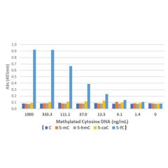
-
anti-5-Hydroxymethylcytosine, Rabbit Monoclonal (RM236)
REV-31-1111-00 REV-31-1111-00-C050 50 µg CHF 623.00
-

-
anti-Crotonyl-Histone H3 (Lys27), Rabbit Monoclonal (RM401)
REV-31-1287-00 REV-31-1287-00-C025 25 µg CHF 261.00REV-31-1287-00-C100 100 µg CHF 581.00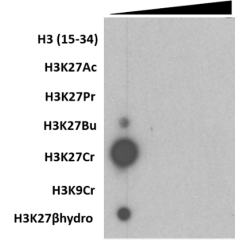
-
anti-Crotonyl-Histone H3 (Lys9), Rabbit Monoclonal (RM339)
REV-31-1225-00 REV-31-1225-00-C025 25 µg CHF 261.00REV-31-1225-00-C100 100 µg CHF 581.00![Western Blot using Anti-Histone H3K9cr Rabbit Monoclonal Antibody RM339 against H3K9cr[Crotonyl-Histone H3 (Lys9)]. Anti-Histone H3 and anti-G6PDH were used as controls. A crotonylation inducing metabolite was used to increase the H3K9cr signal.](https://adipogen.com/media/catalog/product/cache/be9249d907792254897dc476f14d8e2a/r/e/rev-31-1225-00-fig2_wb_h3k9cr_e-300x163.png)
-
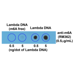
anti-N6-Methyladenosine, Rabbit Monoclonal (RM362)
REV-31-1248-00 REV-31-1248-00-C050 50 µg CHF 477.00