Cookie Policy: This site uses cookies to improve your experience. You can find out more about our use of cookies in our Privacy Policy. By continuing to browse this site you agree to our use of cookies.
AdipoGen Life Sciences
JC-1
As low as
80
CHF
CHF 80.00
In stock
Only %1 left
AG-CR1-3568-M0011 mgCHF 80.00
AG-CR1-3568-M0055 mgCHF 230.00
AG-CR1-3568-50015 x 1 mgCHF 280.00


Specifications / Handling
| Product Details | |
|---|---|
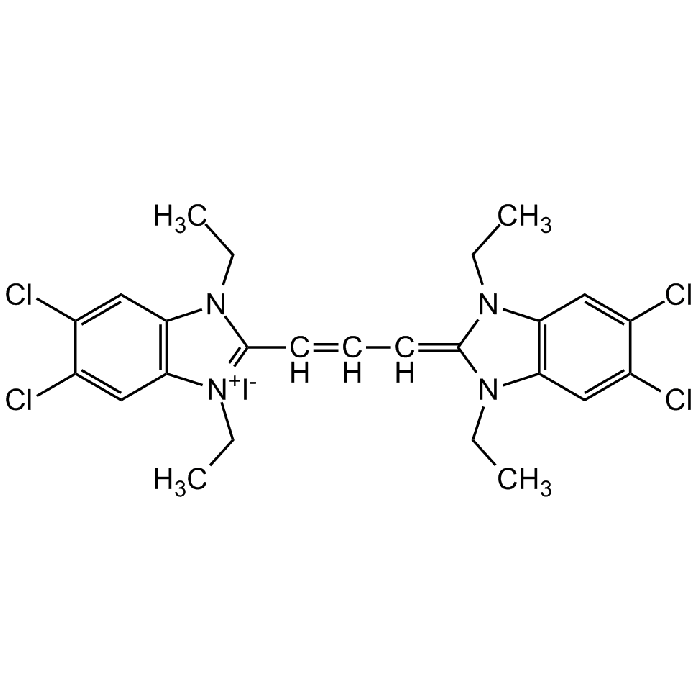
| Synonyms | 5,5',6,6'-Tetrachloro-1,1',3,3'-tetraethylbenzimidazolylcarbocyanine iodide |
| Product Type | Chemical |
| Properties | |
| Formula |
C25H27Cl4IN4 |
| MW | 652.2 |
| CAS | 3520-43-2 and 47729-63-5 |
| Purity Chemicals | ≥98% (NMR) |
| Appearance | Red powder. |
| Solubility | Soluble in DMSO, ethanol, methanol or acetone. |
| Identity | Determined by 1H-NMR and 13C-NMR. |
| Other Product Data |
Application Protocols: See literatures 8, 12 or 13 or the website protocol from Purdue University. |
| InChi Key | FYNNIUVBDKICAX-UHFFFAOYSA-M |
| Smiles | [I-].CCN1C(=CC=CC2=[N+](CC)C3=C(C=C(Cl)C(Cl)=C3)N2CC)N(CC)C2=C1C=C(Cl)C(Cl)=C2 |
| Shipping and Handling | |
| Shipping | AMBIENT |
| Short Term Storage | +4°C |
| Long Term Storage | -20°C |
| Handling Advice |
Keep cool and dry. Protect from light. |
| Use/Stability | Stable for at least 2 years after receipt when stored at -20°C. |
| Documents | |
| MSDS |
 Download PDF Download PDF |
| Product Specification Sheet | |
| Datasheet |
Download PDF |
Scientific Background Information
Product Description
The membrane-permeant dual-emission potential-sensitive JC-1 dye is widely used in apoptosis studies to monitor mitochondrial health by flow cytometry, fluorescence microscopy and in microplate-based fluorescent assays. JC-1 dye can be used as an indicator of mitochondrial membrane potential in a variety of cell types, including myocytes and neurons, as well as in intact tissues and isolated mitochondria. JC-1 accumulates in mitochondria, selectively generating an orange J-aggregate emission profile (590 nm) in healthy cells. After cell injury, as membrane potential decreases, JC-1 monomers are generated, resulting in a shift to green emission (529 nm). The principal advantage of JC-1 relative to other commonly employed fluorescent probes of mitochondrial membrane potential is that it allows qualitative visualization, considering the shift from orange to green fluorescence emission, and quantitative detection, considering the fluorescence intensity ratio.
Product-specific References
- J-aggregate formation of a carbocyanine as a quantitative fluorescent indicator of membrane potential: M. Reers, et al.; Biochemistry 30, 4480 (1991)
- Intracellular heterogeneity in mitochondrial membrane potentials revealed by a J-aggregate-forming lipophilic cation JC-1: S.T. Smiley, et al.; PNAS 88, 3671 (1991)
- A new method for the cytofluorimetric analysis of mitochondrial membrane potential using the J-aggregate forming lipophilic cation 5,5',6,6'-tetrachloro-1,1',3,3'-tetraethylbenzimidazolcarbocyanine iodide (JC-1): A. Cossarizza, et al.; BBRC 197, 40 (1993)
- Mitochondrial membrane potential monitored by JC-1 dye: M. Reers, et al.; Methods Enzymol. 260, 406 (1995)
- JC-1, but not DiOC6(3) or rhodamine 123, is a reliable fluorescent probe to assess delta psi changes in intact cells: implications for studies on mitochondrial functionality during apoptosis: S. Salvioli, et al.; FEBS Lett. 411, 77 (1997)
- Functional assay of multidrug resistant cells using JC-1, a carbocyanine fluorescent probe: J.M. Kühnel, et al.; Leukemia 11, 1147 (1997)
- Flow cytometric measurement of mitochondrial mass and function: a novel method for assessing chemoresistance: M. Mancini, et al.; Ann. Surg. Oncol. 5, 287 (1998)
- Flow cytometric analysis of mitochondrial membrane potential using JC-1: A. Cossarizza & S. Salvioli; Curr. Protoc. Cytom. Chapter 9, Unit 9.14 (2001) (Protocol)
- JC-1: a very sensitive fluorescent probe to test Pgp activity in adult acute myeloid leukemia: O. Legrand, et al.; Blood 97, 502 (2001)
- Mitochondrial and nonmitochondrial reduction of MTT: interaction of MTT with TMRE, JC-1, and NAO mitochondrial fluorescent probes: T. Bernas & J. Dobrucki; Cytometry 47, 236 (2002)
- JC-1, a sensitive probe for a simultaneous detection of P-glycoprotein activity and apoptosis in leukemic cells: D. Chaoui, et al.; Cytometry B Clin. Cytom. 70, 189 (2006)
- Polychromatic analysis of mitochondrial membrane potential using JC-1: E. Lugli, et al.; Curr. Protoc. Cytom. Chapter 7, Unit 7.32 (2007) (Protocol)
- Labeling mitochondria with JC-1: B. Chazotte; Cold Spring Harb. Protoc. 2011, (2011) (Protocol)
- Stable Oxidative Cytosine Modifications Accumulate in Cardiac Mesenchymal Cells from Type2 Diabetes Patients: Rescue by Alpha-Ketoglutarate and TET-TDG Functional Reactivation: F. Spallotta, et al.; Circ. Res. Ahead of print (2017)
- An α-synuclein decoy peptide prevents cytotoxic α-synuclein aggregation caused by fatty acid binding protein 3: N. Fukui, et al.; J. Biol. Chem. 296, 100663 (2021)
- The Atrophic Effect of 1,25(OH)2 Vitamin D3 (Calcitriol) on C2C12 Myotubes Depends on Oxidative Stress: T. Raiteri, et al.; Antioxidants 10, 1980 (2021)
- Aldehyde dehydrogenase 3A1 deficiency leads to mitochondrial dysfunction and impacts salivary gland stem cell phenotype: V. Viswanathan, et al.; PNAS Nexus 1, 1 (2022)
Related Products
-
 JC-10 CDX-T0103
 -
 JC-1 CDX-T0046
 -
 Oxonol V CDX-P0019
 -
 Merocyanin 540 CDX-M0033
 -
 JC-10 (high purity) AG-CR1-3600
 -
 (S)-(+)-Camptothecin AG-CN2-0463