Cookie Policy: This site uses cookies to improve your experience. You can find out more about our use of cookies in our Privacy Policy. By continuing to browse this site you agree to our use of cookies.
PD-1 [CD279]/PD-L1 [CD274] Pathway
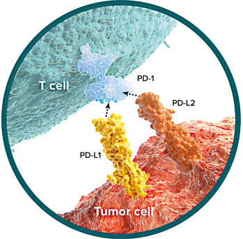 PD-1 (Programmed cell death protein 1; CD279) is a type I transmembrane protein belonging to the CD28/CTLA-4 family of immune receptors. PD-L1 (CD274; B7-H1) and PD-L2 (B7-DC; CD273) are immuno-coinhibitory ligands of the B7 family binding to PD-1. The PD-1/PD-L1 or PD-L2 signaling pathway is a negative regulatory mechanism that inhibits T cell proliferation and cytokine production. Blockade of the PD-1/PD-L1 interaction enhances antitumor immunity. The PD-1 pathway plays a major role in the inhibition of self-reactive T cells and protection against autoimmune diseases. PD-1 and PD-L1 also exist as soluble forms. Elevated levels of soluble PD-1 (sPD-1) are shown in rheumatoid arthritis, skin sclerosis and autoimmune hepatitis. Levels of sPD-L1 are increased in the plasma of cancer patients as well as in cerebrospinal fluid of gliomas. sPD-L1 is a biomarker of poor survival in patients with B cell lymphoma, renal cell carcinoma, metastatic melanoma or lung cancer and is associated with advanced tumor stages.
LIT: PD-1/PD-L1 blockade in cancer treatment: perspectives and issues: J. Hamanishi, et al.; Int. J. Clin. Oncol. 21, 462 (2016) • PD-1/PD-L1 immune checkpoint: Potential target for cancer therapy: F.K. Dermani, et al.; J. Cell Physiol. 234, 1313 (2019)
NEW: PD-1 (human) ELISA Kit - Biomarker for Immuno-Oncology & Autoimmune Diseases
The PD-1 (human) ELISA Kit (Prod. No. AG-45B-0015) is a sandwich ELISA for specific quantitative determination of human soluble PD-1 in serum, plasma and cell culture supernatants. This ELISA Kit shows a very high specificity and sensitivity (1.6pg/ml).
NEW: PD-L1 (human) ELISA Kit - Tumor Biomarker for Poor Survival Prognosis
The PD-L1 (human) ELISA Kit (Prod. No. AG-45B-0016) is a sandwich ELISA for specific quantitative determination of human soluble PD-L1 in serum, plasma and cell culture supernatants. This ELISA Kit shows a very high specificity and sensitivity (0.8pg/ml).
PD-L1 (mouse):Fc (mouse) (rec.) - Biologically Active Proteins
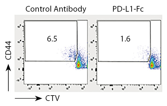
PD-L1 (mouse):Fc (mouse) (rec.) (Prod. No. CHI-MF-110PDL1) is a biologically active recombinant fusion protein that inhibits anti-CD3/CD28 antibody-induced proliferation of mouse T lymphocytes.
| Biological Activity: T cells from C57BL/6 mice were activated with coated anti-CD3/CD28 and PD-L1-Fc or control antibody (10µg/ml). Plots illustrate the percentage of dividing cells by using anti-CD44 and the dye Cell Trace Violet (CTV). . |
 |
UNIQUE anti-PD-1 (mouse), mAb (blocking) (1H10)
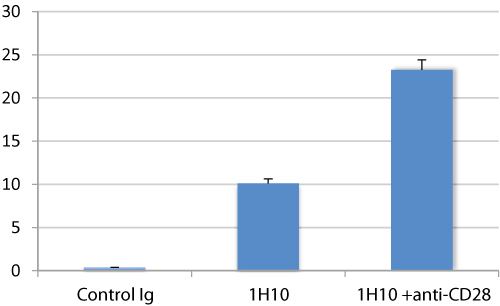
AdipoGen Life Sciences' anti-PD-1 (mouse), mAb (blocking) (1H10) (Prod. No. AG-20B-0075) is a monoclonal antibody that recognizes mouse PD-1 and works specifically in Flow Cytometry and Functional Application. The antibody blocks PD-1 binding and induces a rapid activation and proliferation of T cells at concentration of 0.25µg/2x105 cells. The antibody is available with or without preservatives.
|
Figure: PD-1 receptor-induced CD4 T cell activation and proliferation by PD-1 (mouse), mAb (blocking) (1H10) (AG-20B-0075).
|
 |
NEW IHC-Competent Antibodies for PD-1 and PD-L1 Staining
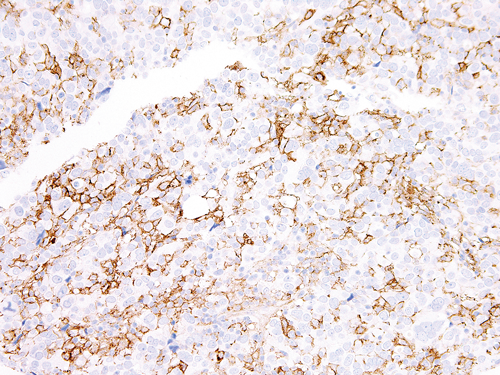
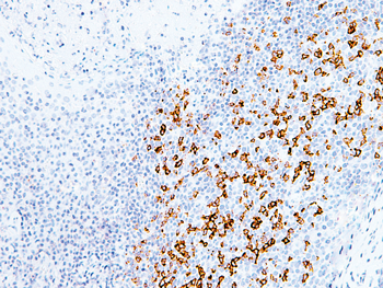
AdipoGen Life Sciences' anti-PD-1 (human), mAb (AG-IHC001) (Prod. No. AG-20B-6020) and anti-PD-L1 (human), mAb (AG-IHC411) (Prod. No. AG-20B-6022) are monoclonal antibodies that recognize human PD-1 and PD-L1, respectively, and work specifically in Immunohistochemistry Application. The antibodies are available in catalog and BULK sizes.
|
Figure: IHC Staining of PD-1 in human tonsil tissue using anti-PD-1 (human), mAb (AG-IHC001) (Prod. No. AG-20B-6020) (left side).
Figure: IHC Staining of PD-L1 in human lung tissue using anti-PD-L1 (human), mAb (AG-IHC411) (Prod. No. AG-20B-6022) (right side).
|
 ; ; |
Biologically Active PD-1 and PD-L1 Proteins
| Product Name | PID | Source | Endotoxin | Species |
| PD-1 (mouse):Fc (mouse) (rec.) | CHI-MF-110PD1 | CHO cells | <0.06EU/µg | Mouse |
| PD-1 (mouse):Fc (human) (rec.) | CHI-MF-111PD1 | HEK 293 cells | <0.06EU/µg | Mouse |
| PD-1 (human) (rec.) (untagged) | CHI-HF-200PD1 | HEK 293 cells | <0.01EU/µg | Human |
| PD-1 (human) (rec.) (His) | CHI-HF-201PD1 | HEK 293 cells | <0.01EU/µg | Human |
| PD-1 (human):Fc (human) (rec.) | CHI-HF-210PD1 | CHO cells | <0.06EU/µg | Human |
| PD-1 (human):Fc (human) (rec.) (non-lytic) | CHI-HF-220PD1 | CHO cells | <0.06EU/µg | Human |
| PD-1 (human):Fc (mouse) (rec.) | CHI-HF-211PD1 | HEK 293 cells | <0.005EU/µg | Human |
| PD-L1 (mouse):Fc (mouse) (rec.) (non-lytic) | CHI-MF-120PDL1 | CHO cells | <0.06EU/µg | Mouse |
| PD-L1 (human) (rec.) (untagged) | CHI-HF-200PDL1 | HEK 293 cells | <0.01EU/µg | Human |
| PD-L1 (human) (rec.) (His) | CHI-HF-201PDL1 | HEK 293 cells | <0.01EU/µg | Human |
| PD-L1 (human):Fc (human) (rec.) | CHI-HF-210PDL1 | CHO cells | <0.06EU/µg | Human |
| PD-L1 (human):Fc (human) (rec.) (non-lytic) | CHI-HF-220PDL1 | CHO cells | <0.06EU/µg | Human |
| PD-L1 (human):Fc (mouse) (rec.) | CHI-HF-211PDL1 | HEK 293 cells | <0.005EU/µg | Human |
| PD-L2 (mouse):Fc (mouse) (rec.) | CHI-MF-110PDL2 | CHO cells | <0.06EU/µg | Mouse |
| PD-L2 (human):Fc (human) (rec.) | CHI-HF-210PDL2 | CHO cells | <0.06EU/µg | Human |
| PD-L2 (human):Fc (human) (rec.) (non-lytic) | CHI-HF-220PDL2 | CHO cells | <0.06EU/µg | Human |
| PD-L2 (human):Fc (mouse) (rec.) | CHI-HF-211PDL2 | CHO cells | <0.06EU/µg | Human |
| Other PD-1 and PD-L1 Proteins |
VALIDATED Antibodies for PD-1 and PD-L1 Research
| Product Name | PID | Isotype | Applications | Species |
| PD-1 (mouse), mAb (blocking) (1H10) (PF) | AG-20B-0075PF | Rat IgG2aκ | FACS, FUNC | Mouse |
| PD-1 (human), mAb (ANC4H6) |
ANC-279-020 |
Mouse IgG1κ | FACS | Human |
| PD-1 (human), mAb (AG-IHC001) | AG-20B-6020 | Mouse IgG1κ | IHC, | Human |
| PD-L1 (human), mAb (ANC6H1) |
ANC-274-020 |
Mouse IgG1κ | FACS | Human |
| PD-L1 (human), mAb (AG-IHC411) | AG-20B-6022 | Mouse IgG1κ | IHC, | Human |
| PD-L2 (human), mAb (ANC8D12) |
ANC-273-020 |
Mouse IgG2aκ | FACS, FUNC | Human |
|
More Information |
|
Downloadable Flyer |
|
PD-1/PD-L1 Signaling Pathway Product Flyer Released - September 2025 |
Download |
